
Headaches from neck pain: should physical therapy for cervicogenic headaches look beyond the cervical spine?
Can posture and shoulder blade position drive headaches from neck pain? We break down the research and what it means for your treatment.

Headaches from neck pain are one of the most frustrating presentations in the clinic. Cervicogenic headache physical therapy has traditionally focused on the cervical spine — and for good reason. Manual therapy to the cervical joints works, and exercises targeting the deep neck flexors help too. The research supports both.
But what if the neck isn't the whole story?
A 2005 case report published by McDonnell, Sahrmann, and Van Dillen explored whether impairments in the scapulothoracic and lumbar regions — not just the cervical spine — could be contributing factors in cervicogenic headache. More specifically, they asked whether treating those regions could meaningfully reduce a patient's headache frequency and intensity.
The study
This was a case report, meaning it describes the examination and treatment of a single patient. Case reports sit at the lowest level of the evidence hierarchy — they cannot establish cause and effect, and their findings cannot be generalized. With that context in mind, this paper is best read as a clinical reasoning framework rather than proof of efficacy.
The patient was a 46-year-old male with a 7-year history of cervicogenic headache. His average pain was 5 out of 10, with peaks of 10 out of 10, and he reported constant symptoms. His Neck Disability Index score was 31 out of 50, placing him in the severe disability category. His symptoms were aggravated by using his arms — whether working with horses or sitting at a computer — and he frequently woke up at night from headache pain.
One important detail: one week before starting physical therapy, he received trigger point injections to the posterior cervical and upper trapezius region. These provided 24 to 48 hours of complete pain relief, and his pain had dropped to 3 out of 10 by the time he arrived for his first PT visit.
The examination
The treating therapist — working within Sahrmann's Movement Impairment Syndrome framework — assessed the cervical, scapulothoracic, and lumbar regions together rather than in isolation.
Key findings included forward head posture with excessive upper cervical extension, marked scapular abduction and depression bilaterally, thoracic kyphosis, and significant weakness of the middle and lower trapezius, rhomboids, and lower abdominals. The deep neck flexors were so weak that formal strength testing wasn't even possible at baseline.
One of the more clinically interesting findings came from a simple test: the examiner manually elevated and adducted the patient's scapulae while he performed cervical rotation. His range of motion increased by 10 degrees in both directions and his headache pain decreased. This suggested that scapular positioning was directly influencing his cervical symptoms — and became the basis for treatment.
The intervention
The patient was seen 7 times over 3.5 months. No manual therapy was used at any point.
Treatment focused entirely on active exercise and functional modification. The exercise program targeted abdominal strength and control, deep neck flexor activation, scapular retraction and elevation, and full shoulder range of motion without compensatory neck or lumbar movement. Functional instruction emphasized supporting the weight of the arms throughout the day — resting forearms on a desk, placing hands in pockets while standing — to reduce the constant downward pull on the cervical spine.
The results
The results across those 7 visits were notable.
By the fourth visit — just 25 days in — the patient was going several days at a time without any headache. By the seventh visit, headaches had dropped to once per week at an intensity of 1 out of 10. His NDI score fell from 31 to 11, shifting from severe to mild disability. Cervical rotation improved from 39 degrees (painful) to 50 degrees (pain-free), and cervical extension went from 25 degrees (painful) to 40 degrees (pain-free). Scapular position improved from 17.8 cm lateral to 11.4 cm lateral from midline.
At a 5-month follow-up phone call, he was sleeping through the night without medication, performing all activities of daily living, and managing flare-ups independently with his exercises.
Limitations of the study:
This is a single case report. There is no control group, no blinding, and no way to determine whether improvement was due to the intervention, natural history, regression to the mean, or the trigger point injections administered the week prior. The trigger point injection in particular is a notable confound — the patient was already improving before PT began.
No independent outcome assessor. The treating therapist was also involved in measuring and reporting outcomes, which introduces the potential for bias.
The primary outcomes — headache frequency and intensity — relied on patient self-report without a structured diary or validated headache frequency tool.
The passive correction of scapular position test used in the examination has not been formally validated as a diagnostic tool.
With only one patient, there is no way to know which components of the intervention — the scapular exercises, the lumbar work, the abdominal training, the functional modifications — were actually driving the improvement, or whether any of them were.
Clinical implications for cervicogenic headache physical therapy
At first glance, the results are compelling. A patient with 7 years of constant, debilitating headaches from neck pain improved dramatically with just 7 visits — and without a single manual therapy technique.
But the more important takeaway may be the clinical reasoning behind the approach.
The idea that scapular depression and abduction can transfer the weight of the upper extremities through the cervicoscapular muscles onto the posterior cervical spine is biomechanically plausible. The idea that lumbar extension drives compensatory cervical extension throughout the day is similarly logical. These are not novel concepts — regional interdependence has been discussed extensively in the PT literature — but applying them systematically to cervicogenic headache is less common in practice.
The passive correction of scapular position test is particularly worth noting. It takes seconds to perform and gives the clinician immediate feedback about whether scapular mechanics are contributing to cervical symptoms. If range of motion improves and pain decreases with manual scapular support, that's a meaningful clinical signal.
What this study cannot tell us is whether adding scapulothoracic and lumbar treatment to a standard cervical approach actually improves outcomes. That question has not been answered in a controlled trial. And given that manual therapy to the cervical spine has a solid short-term evidence base for CH, it is also worth asking: would this patient have done just as well — or better — with the addition of manual therapy?
Final thoughts
This is not a study you should use to change your protocol. It is something that should prompt you to look further than the cervical spine when you assess a patient with headaches from neck pain.
The cliche “shoulders down and back” is not always the answer. In this case, “shoulders down”, or scapular depression, may actually exacerbate cervicogenic symptoms.
Are the scapulae sitting low and wide? Is the patient extending through the neck every time they raise their arm? Is there a lumbar alignment that is feeding into prolonged cervical extension posture throughout the day? These are questions worth asking. The evidence for acting on the answers is still limited — but the clinical logic is sound.
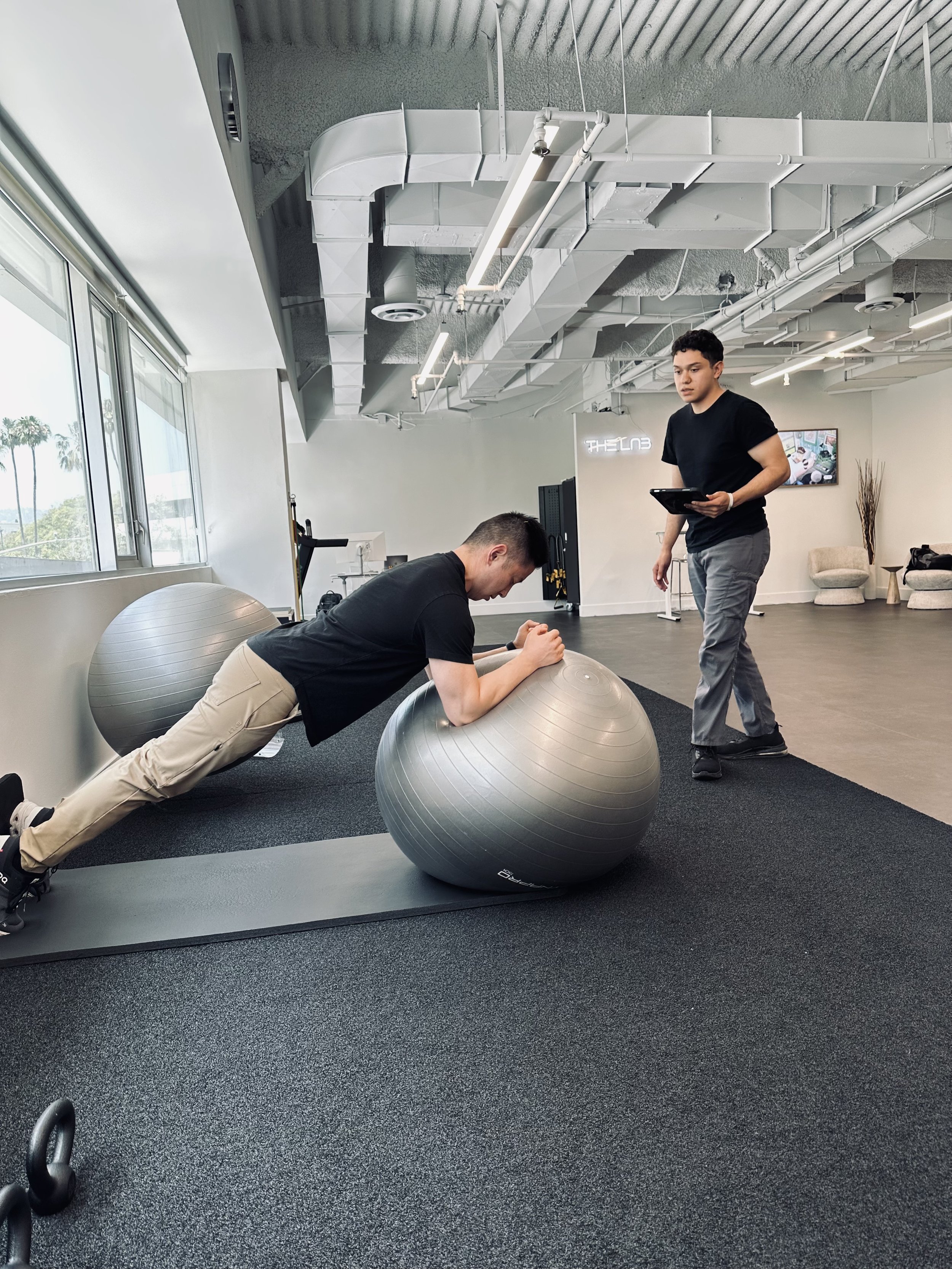
At The LAB Doctors of Physical Therapy, we take a whole-body approach to cervical spine conditions, building treatment programs grounded in both the evidence and the individual in front of us. Click here to learn more about our services.
Citations
1.) McDonnell MK, Sahrmann SA, Van Dillen L. A specific exercise program and modification of postural alignment for treatment of cervicogenic headache: a case report. J Orthop Sports Phys Ther. 2005;35(1):3–15.
Should you treat the hip for low back pain?
Does treating the hip improve low back pain? A 2021 randomized trial shows no added benefit. Learn what actually drives recovery and effective physical therapy.

Low back pain continues to be one of the most common reasons patients seek physical therapy. The more concerning issue is that non-specific low back pain, which accounts for approximately 90% of all low back cases, is widely under diagnosed due to the complexity of the region. Once red flags and specific mechanisms of injury are excluded, any combination of musculoskeletal, neurological and psychosocial factors can elicit symptoms in the low back.
Recently, there has been a growing emphasis on the role of the hip, particularly when patients present with limited mobility, weakness or asymmetries. In the clinical setting, it is now common to hear that “tight hips are causing your back pain”, or “weak glutes are the problem.”
But an important question remains.
Does treating the hip actually improve outcomes in patients with low back pain?
A 2021 randomized controlled trial published by Burns et al set out to answer this directly.
The study
This randomized controlled trial study evaluated whether adding hip-specific treatment to standard low back physical therapy led to better outcomes.
Participants were 76 adults with 1) low back pain and 2) concurrent hip impairments, which included mobility and/or strength deficits of the hip. These participants were then divided into two groups:
Group 1 was given Lumbar Treatment Only (LBO).
Group 2 was given both Lumbar + Hip Treatment (LBH).
Both groups received standard physical therapy for the low back, including 1) exercise, 2) manual therapy and 3) patient education.
The hip-treatment group received additional interventions, including 1) hip strengthening, 2) mobility work and 3) manual therapy targeting the hip.
It is important to note that the lumbar treatment was not strictly standardized—it was based on clinician judgment, reflecting real-world physical therapy practice.
Outcome measures included 1) pain (Numeric Pain Rating Scale), 2) disability (Oswestry Disability Index), 3) fear-avoidance beliefs, and 4) functional improvement.
Patient follow-up was obtained at baseline, 2 weeks, discharge, 6 months and 12 months post-intervention.
The results
Both LBO and LBH groups improved significantly over time, but there was no additional benefit from hip treatment.
In other words, there were no meaningful differences between groups at any time point.
The interesting finding, however, was that the group receiving additional hip treatment had higher fear-avoidance beliefs at 2 weeks and discharge. This subtlety may suggest that additional treatment may not necessarily improve patient confidence or outcomes.
Limitations of the study: 1) variability in treatment approaches. The details of low back treatment were not included, and it was based on therapist discretion to mimic real-world applications. However, without standardization of what low back care entailed, we do not know whether there were inadvertently hip-involved interventions used within the LBO group. 2) The study was not blinded, meaning that patients and therapists knew which group they were placed under. This increased the risk of both observer and participant expectation bias, which could vastly affect the results. 3) The education aspect of care, while directed to be centered around the low back, may have also included the hip. 4) Small sample size and missing data at follow-up.
Clinical implications for physical therapists
At first glance, the study’s findings challenge the common assumption that, “if the hip is impaired, treating it will improve low back pain.” Rather yet, the study suggests that identifying and addressing a hip impairment does not necessarily benefit low back pain, and that improvements may be driven more by general movement, load tolerance and gradual return to activity.
The conclusions of the study suggest that, for most patients with low back pain, focusing treatment on the spine alone may be sufficient—even when hip deficits are present. But were the participants in the “low back only” group truly receiving treatment exclusive to the low back? Learning lumbopelvic awareness through a cat-cow exercise will also affect movements in the hip. Education on protection of the low back includes promoting movement in the hip to offload the spine. Even without targeted hip interventions, it is unlikely the hip was truly ignored. Unless we know that the experiment’s “low back only” group was truly limited to the lumbar spine (ie. core strengthening, spinal mobilizations) we should be cautious to jump to the conclusion that focusing on spine alone suffices.

Nonetheless, the study does provide standard procedures for the participants who received additional hip treatment. This included specific manual therapy procedures and a progressive exercise protocol for the hip. And even so, this group did not perform better than the other, strengthening the conclusion that adding hip-specific work does not necessarily provide additional benefit.
The focus is then as such: More is not always better. The goal is precision, not excess. Overcomplicating care can reduce clarity for the patient and shift focus away from meaningful progress. Therefore, while a patient can indeed have “tight hips”, “weak glutes”, or “muscle imbalances”, these are not always the primary drivers of pain. Training hip stability and mobility have almost become synonymous with low back pain protocols, and it’s important that we take a step back and acknowledge the distinction between the two.
Final thoughts? This study reinforces an important yet forgotten principle in rehabilitation: effective treatment is not about doing more—it’s about doing what matters most.
At The LAB Doctors of Physical Therapy, we build an evidence-based treatment program that is both effective and efficient. Click here to learn more about our services.
Citations
1.) Burns SA, Cleland JA, Rivett DA, O’Hara MC, Egan W, Pandya J, Snodgrass SJ. When treating coexisting low back pain and hip impairments, focus on the back: adding specific hip treatment does not yield additional benefits: a randomized controlled trial. J Orthop Sports Phys Ther. 2021;51(12):581-601.
Proprioception vs Strengthening: what works best for rotator cuff–related shoulder pain?
A 2025 randomized clinical trial shows strengthening exercises alone are just as effective as adding proprioception for treating rotator cuff–related shoulder pain.

What is rotator cuff-related shoulder pain?
The rotator cuff is comprised of four muscles (supraspinatus, infraspinatus, teres minor and subscapularis), whose primary role is to provide stability to the shoulder joint. Let’s put it this way: your shoulder joint is inherently the most unstable joint in the body, as it’s a ball-and-socket joint that is analogous to a golf ball on a tee. With that said, these four muscles of the rotator cuff must be not only be strong individually, but also as a unit that coordinates as a team to provide both static and dynamic stability to the shoulder as you move your arm about. Injury to one or more of these muscles can affect your arm’s ability to move in a bio-mechanically sound manner, translating to pain with movement over time.
What’s the difference between proprioception and strengthening?
Proprioception is your body’s ability to perceive joint awareness in space. Your central nervous system processes somatosensory input from receptors in your joints to produce motor output. This, for example, would allow you to catch yourself from rolling your ankle on some unforeseen rock while hiking on a trail.
Strengthening, on the other hand, is providing a resistive stimulus that promotes increased force production through adaptation. When your muscle is physically taxed repeatedly over time, it will adapt and grow stronger to conform to demands.
Research has shown favorable outcomes with incorporating both proprioceptive and strength training for patients with osteoarthritis or ankle sprains. This following study looks to see whether the same applies for rotator cuff-related shoulder injuries, which may shine some light on the most efficient physical therapy protocols for shoulder rehab.
The study
Buccioli et al in this 2025 study looks at whether there are any advantages of a combinatory program of proprioceptive and strength training versus strength training alone for subjects with rotator cuff-related shoulder pain (RCRSP). The study was a randomized controlled trial with two primary groups:
Group 1 was given strengthening exercises alone (the control).
Group 2 was given strengthening exercises plus proprioceptive training (the experimental).
Both groups underwent training over the course of 8 weeks, with data collected at start of treatment, followed by 2 months and 5 months from start of intervention.
Outcome measures included Shoulder Pain and Disability Index (SPADI), pain (Visual Analog Scale), range of motion (ROM), isometric strength, and joint position sense (JPS).
The results
Both groups improved significantly in all outcomes measures, including SPADI, pain, ROM, strength and joint position sense
Limitations: 1) The study didn’t include a non-intervention control group, which would have been nice to see whether the favorable outcome measures were indeed due to the allocated intervention versus just the natural healing progression of time. 2) The same physical therapist was used to treat both groups, increasing the risk for performance bias. However, since the hypothesis that proprioceptive training would yield better outcome measures was disproved based on the data of this study, it’s likely this bias was controlled to some degree. 3.) The 5-month follow-up had a 26% decreased follow-up in the control group, and 29% in the experimental group.
Clinical implications for physical therapists
For a more efficient treatment approach, it may suffice to focus on the strength training rather than dividing time between strength and proprioceptive training. By all means, though, this doesn’t undermine the importance of proprioceptive training. Rather yet, enhanced proprioception may occur while performing strength training, so it may be a two-birds-one-stone scenario. Put it this way: changes in motor output following proprioceptive exercise may either result from enhanced somatosensory information (which in turn improved motor output) or improved sensorimotor integration. Since the data from both groups shows increased proprioception post-intervention, it is possible that shoulder strengthening exercises alone can improve sensorimotor integration, which increases proprioception.
Citations
1.) Buccioli V, van de Water A, van den Noort JC, et al. Proprioceptive exercises combined with strengthening exercises are not superior to strengthening exercises alone for shoulder pain and disability in individuals with chronic rotator cuff-related shoulder pain: a randomized controlled trial. J Orthop Sports Phys Ther. 2025;55(7):495–511. https://www.jospt.org/doi/10.2519/jospt.2025.13097.
2.) Aman JE, Elangovan N, Yeh IL, Konczak J. The effectiveness of proprioceptive training for improving motor function: a systematic review. Front Hum Neurosci. 2015;8:1075. https://doi.org/10.3389/fnhum.2014.01075.
Ankle inversion sprains: the added benefits of manual therapy combined with stabilization exercises
This article looks at a study highlighting the benefits of a multi-modal approach to addressing ankle inversion sprains.

Ankle inversion sprains occur commonly in physical active people, with an annual incidence rate of 7 injuries per 1000 people. It occurs when the foot rolls inward, causing damage to the ligaments on the outer portion of the ankle. Not only does this cause pain and limited tolerance to everyday activities, but damage to ligaments can negatively impact proprioception, or awareness of joint positioning, which can decrease performance in balance and set the stage for recurrent injuries.
The following study compares the effects of manual therapy with exercise versus exercise alone, when treating an ankle inversion sprain.
The study:
Cleland et al performed a randomized controlled trial in 2013, where patients with ankle inversion injuries underwent a four-week intervention program. The study randomly allocated subjects into two groups:
Group 1 was given exercise alone.
Group 2 was given the same exercise program as Group 1, plus a manual therapy regime. The manual therapy regime consisted of mobilizations to the proximal and distal tibiofibular joints, talocrural and subtalar joints.
Outcome measures were the Foot and Ankle Ability Measures (ADL and Sports), Lower Extremity Functional Scale (LEFS), Numeric Pain Rating Scale (NPRS) and Global Rating of Change (GRC). Post-intervention measures were taken after the 4-week intervention and at 6-months.
The results:
The overall group-by-time interaction for the mixed-model analysis of variance was statistically significant for the FAAM activities of daily living subscale (P<.001), FAAM sports subscale (P<.001), Lower Extremity Functional Scale (P<.001), and pain (P⩽.001). Improvements in all functional outcome measures and pain were significantly greater at both the 4-week and 6-month follow-up periods in favor of the MTEX group.
There was no statistically significant difference in the recurrence of ankle sprain rate between the both groups.
Limitations of the study: 1.) subjects were not blinded to group assignment, 2.) all outcome measures were questionnaires, and there were no objective outcome measures, such as improvements in range of motion, strength, 3.) there was no control group, so improvements cannot be attributed to the treatment intervention or that of placebo effect, and 4.) participants of Group 2 (exercise + manual therapy) spent more time with a physical therapist, which may encourage bias.
What this means:
According to the results of this study, a multimodal approach integrating both ankle stability exercises and manual therapy improves pain and increased tolerance to activities of daily living and return to sport. The nature of improvement is still unknown, and there are several explanations that may account for results favoring manual therapy intervention:
1.) manual therapy may help restore motion in the joints, consequently improving function and decreasing pain. This is the primary intention of manual therapy.
2.) the effects of manual therapy may be neurophysiological. That is, it may play a role in undoing a disrupted neural feedback system to the dynamic stabilizers of the ankle via stimulation of mechanoreceptors in the ankle.
3.) manual therapy may result in reduction of inflammatory cytokines, an increase in beta endorphins, and hypoalgesia.
No matter the case, manual therapy seems to play a role in improving pain and perceived tolerance to activities of daily living. Below are the manual therapy techniques used in the appraised study.
REFERENCES:
Cleland JA, Mintken P, McDevitt A, Bieniek M, Carpenter K, Kulp K, Whitman JM. Manual Physical Therapy and Exercise Versus Supervised Home Exercise in the Management of Patients With Inversion Ankle Sprain: A Multicenter Randomized Clinical Trial. Journal of Orthopaedic & Sports Physical Therapy. 201343:7,443-455
Shoulder impingement: exercise versus exercise and manual therapy
What is the best course of action for shoulder pain caused by subacromial impingement? This article compares exercise alone versus exercise and manual therapy on addressing this particular issue.

Shoulder impingement syndrome (SIS) is a common problem derived from repetitive use of the arm at or above shoulder level. While there is proven success on the management of SIS through conservative treatment, researchers and physical therapists still debate the best course of action to most efficiently address this problem.
The study:
Camargo et al performed a randomized controlled trial in 2015, comparing two approaches in managing patients with SIS:
Group 1 was given exercise alone.
Group 2 was given the same exercise program as Group 1, plus a manual therapy regime.
Outcome measures included pain, mechanical sensitivity, and changes in scapular kinematics. Post-intervention measures were taken after the 4-week intervention.
The results:
No significant changes in scapular kinematics were found with both groups pre- and post-intervention.
Both groups experienced significant decreases in pain at rest and during movement when comparing pre- and post-intervention, but had no significant differences between groups.
Both groups experienced increased mechanical sensitivity when comparing pre- and post-intervention, but had no significant differences between groups.
Limitations of the study: 1.) participants did not have acute SIS, and therefore the results do not pertain to a more recent flare-up of SIS, and 2.) no long-term follow-up, as results were only taken after the 4-week intervention.
What’s most noteworthy of this study, however, is that despite improvements in pain and mechanical sensitivity, scapular kinematics have not significantly changed even after intervention. This suggests that improvements in pain and mechanical sensitivity cannot be explained by changes in scapular motion.
What this means:
With respect to this study, the addition of manual therapy did not provide any additional benefit in improving scapular kinematics, pain levels or mechanical sensitivity for patients with shoulder impingement syndrome. This suggests that exercise alone may suffice in addressing the issue. Segueing to some helpful exercises below:
REFERENCES:
Camargo PR, Alburquerque-Sendín F, Avila MA, Haik MN, Vieira A, Salvini TF. Effects of Stretching and Strengthening Exercises, With and Without Manual Therapy, on Scapular Kinematics, Function, and Pain in Individuals With Shoulder Impingement: A Randomized Controlled Trial. J Orthop Sports Phys Ther. 2015 Dec;45(12):984-97. doi: 10.2519/jospt.2015.5939. Epub 2015 Oct 15. PMID: 26471852.
Carpometacarpal (CMC) osteoarthritis: thumb pain and the role of physical therapy
Carpometacarpal osteoarthritis are among one of many causes of thumb pain. This randomized controlled trial looks at the benefits of exercise and manual therapy on this problem.

Thumb pain has never been a more common problem than it has been now. Computers and smart devices aren’t going away anytime soon, and neither will the incessant demand on our text-punching, mouse-guiding thumbs. Over time, repetitive overuse of thumbs can lead to degenerative changes, such as osteoarthritis of the thumb’s base joint.
The study:
Villafane et al performed a randomized controlled trial consisting of 60 patients diagnosed with 1st carpometacarpal osteoarthritis. These cases were divided into 2 groups:
The experimental group (EG) received a multimodal treatment approach, consisting of exercise and manual therapy (joint and neural mobilizations).
The control group (CG) received a sham intervention, which consisted of pulsed ultrasound at 0 W/cm2 applied on the base of the thumb.
Both groups underwent 12 sessions of treatment over 4 weeks. Outcome measures included reported pain, pinch and grip strength, and pain pressure threshold over 1st CMC, scaphoid and hamate bones.
The results:
The EG reported significant decrease in pain levels from pre-intervention (5.0) to post-intervention (1.9), which continued to decrease at 1-month and 2-month follow-up (1.5), while the CG had smaller decrease from pre-intervention (5.0) to post-intervention (4.9). p<0.001.
There were no statistically significant changes in outcome measure of pinch/grip strength and pain pressure threshold in both EG and CG when comparing pre-intervention to post-intervention.
Limitations in this study: 1.) sample size too small. 2.) the subjects did not complain of grip/pinch weakness pre-intervention, and therefore the prescribed exercises may not have had a strengthening effect. 3.) no measure of function was included as an outcome measure. 4.) no long-term follow-up after 2 months.
What this means:
A multimodal treatment approach including manual therapy and exercise can help significantly improve pain associated with osteoarthritis of the thumb joint. Though this study included only patients with osteoarthritis, it is curious whether similar treatment procedures may be utilized to manage other diagnoses involving the thumb. Further research is needed to help confirm this hypothesis.
The vid below shows some examples of thumb pain treatment used in this article’s experiment.
REFERENCES:
Villafañe JH, Cleland JA, Fernández-de-Las-Peñas C. The effectiveness of a manual therapy and exercise protocol in patients with thumb carpometacarpal osteoarthritis: a randomized controlled trial. J Orthop Sports Phys Ther. 2013 Apr;43(4):204-13. doi: 10.2519/jospt.2013.4524. Epub 2013 Mar 13. PMID: 23485660.
Maternal weight gain and gestational diabetes: exercise during pregnancy
Excessive maternal weight gain and gestational diabetes mellitus are among a number of conditions that can occur during pregnancy. This article looks at a study analyzing the effects of exercise during prenatal care.

The human body undergoes significant changes during pregnancy and delivery, which can ultimately affect maternal health and fetal well-being. Healthcare professionals have linked excessive maternal weight gain with adverse conditions, such as gestational diabetes—a condition where blood sugar levels become too high and is diagnosed for the first time during pregnancy.
The study:
Barakat et al performed a randomized controlled study analyzing the effects of exercise during pregnancy. 456 participants were allocated into two groups:
Control group (CG): 222 participants received obstetric standard care from healthcare professionals.
Experimental group (EG): 234 participants participated in a supervised, moderate exercise program 3 times a week (50-55 min/session) from 8-10 weeks to 36-38 weeks gestation. This exercise program consisted of warm-up, aerobic exercise, mild resistance training, coordination and balance, stretching, pelvic floor strengthening, and relaxation.
Outcome measures included maternal weight gain, excessive gestational weight gain categorization (based on 2009 Institute of Medicine guidelines using pre-pregnancy BMI), oral glucose tolerance test (OGTT) and cases of gestational diabetes mellitus (GDM). Weight measures were obtained on the first prenatal consult and last clinical visit (~week 36-38), while OGTT and GDM were collected from hospital records during weeks 24-26.
The results:
Maternal weight gain was significantly lower in the EG versus CG (12.19 kg vs 13.33 kg, respectively).
Excessive weight gain was significantly higher in CG versus EG (30.2% vs 25.5%, respectively)
Significant differences were also noted in OGTT tests (EG = 116.56 vs CG = 121.63 mg/dL)
The ratio of women diagnosed with GDM was higher in the CG than the EG (6.8% vs 2.6%, respectively)
Limitations in the study: 1) lack of information regarding nutritional intake of participants, and 2) participants were chosen from two hospital locations in Spain, which may affect its external validity. The study’s main strength, however, was the number of participants who have completed the study from start to finish.
What this means:
The study suggests that exercise of at least three times a week can help lower excessive maternal weight gain and act as a preventative for secondary conditions such as gestational diabetes mellitus. Therefore, the results of the study were largely in favor of exercise and its positive effects on maternal health.
Most people ask whether it’s okay to exercise, particularly in the later stages of pregnancy. This study suggests yes, we do—but with supervision.
REFERENCES:
Barakat R, Refoyo I, Coteron J, Franco E. Exercise during pregnancy has a preventative effect on excessive maternal weight gain and gestational diabetes. A randomized controlled trial. Braz J Phys Ther. 2019 Mar-Apr;23(2):148-155. doi: 10.1016/j.bjpt.2018.11.005. Epub 2018 Nov 17. PMID: 30470666; PMCID: PMC6428908.
Hip impingement: hip-targeted exercise versus hip-targeted exercise with trunk stabilization
Femoroacetabular hip impingement (FAI), best described as a pinching anterior hip pain. This article looks at another means of addressing the problem on top of the standard hip strengthening protocol.

Femoroacetabular impingement occurs when the femur repetitively abuts the rim of its socket (acetabulum), resulting in front hip pain, with long-term complications including possible labrum damage and osteoarthritis. The standard protocol has included stretching and strengthening the hip musculature to minimize this abutting; this article looks at a study that also incorporates trunk strengthening.
The study:
Aoyama et al performed a prospective, randomized controlled trial looking at 20 female participants with hip impingement, more specifically the effects of adding trunk stabilization exercises on top of a hip strengthening protocol.
The control group received the hip and pelvic girdle protocol, which consisted of gluteus medius/maximus strengthening and lumbopelvic awareness exercises.
The trunk stabilization exercise group received the same hip and pelvic girdle protocol, as well as additional exercises targeting the transverse abdominis and oblique musculature.
Outcome measures were recorded prior to treatment, at 4 weeks post-intervention and 8 weeks post-intervention. Measures included hip range of motion, strength and patient-reported pain levels.
The results:
The trunk group had significantly improved hip flexion range of motion at 4-wks post-intervention, compared to the control group.
The trunk group had significantly improved hip abduction strength at 4-wks post-intervention, with no significant improvement in the control group.
Patient-reported outcomes (iHOT12 and Vail hip scores) were significantly improved in both groups at 8-weeks post-intervention, though the trunk group had greater improvement than the control group.
What this means:
The primary limitations of this study included its small sample size (n=20) and all-female participant pool, which may compromise its applicability to a broader audience.
Regardless, the most noteworthy take-aways from the study’s results were the improvements in hip range of motion and strength at 4-wks post-intervention, which suggests that adding a trunk stabilization program, along with hip strengthening, can boost short-term outcomes.
REFERENCES:
Aoyama M, Ohnishi Y, Utsunomiya H, Kanezaki S, Takeuchi H, Watanuki M, Matsuda DK, Uchida S. A Prospective, Randomized, Controlled Trial Comparing Conservative Treatment With Trunk Stabilization Exercise to Standard Hip Muscle Exercise for Treating Femoroacetabular Impingement: A Pilot Study. Clin J Sport Med. 2019 Jul;29(4):267-275. doi: 10.1097/JSM.0000000000000516. PMID: 31241527; PMCID: PMC6613832.
Cubital tunnel syndrome: reviewing our conservative options
Leaning on your elbows too much can mean bad news, such as the onset of cubital tunnel syndrome. This week, we look at the conservative options available that address this condition.

With no promise of returning back to the office anytime soon, The LAB is continuing to see more problems associated with working from home, including cubital tunnel syndrome. Considered the second most common neuropathy of the upper extremity (first being carpal tunnel syndrome), cubital tunnel syndrome is caused by compression and irritation of the ulnar nerve, due to direct pressure on the inner elbow (near the “funny bone” region) or repetitive, maximal stretching into elbow flexion. This includes resting the elbows on a hard surface for long hours at a time.
The study:
Kooner et al performed a systematic review that looked at all conservative options for treating cubital tunnel syndrome. Twenty studies were included for analysis, with the most common outcome measures being patient-reported outcomes and nerve conduction studies. The average treatment duration was 3-months, with most intervention methods including education/activity modification, splinting, steroid/lidocaine injection, nerve mobilization/gliding, ultrasound, laser therapy, NSAIDs administration and physical therapy.
The results:
Two studies showed that activity modification and patient education alone resolved symptoms of 44-66% of patients after one year.
One study, a randomized controlled trial, compared activity modification/patient education with splinting and nerve gliding techniques. There were no significant differences between both groups, yet 90% of all participants demonstrated clinical improvement at 6-months.
One prospective cohort study looked at the effects of patients receiving splinting and activity modification over a 3-month period. Participants demonstrated improved questionnaire scores (DASH) and grip strength; 82% of patients became symptoms-free over a 2-year period.
One study showed no significant difference between steroid injections and placebo, with only 30% success rate for treatment.
Due to lack of heterogeneity, the systematic review had difficulty grouping which specific interventions trumped the others. There was also a lack of quality studies, and sample sizes were too small.
What it means:
Based on the studies, the author suggests that activity modification, patient education and splinting all prove to be among the most effective conservative means of addressing cubital tunnel syndrome.
Below are examples of exercises that also address cubital tunnel syndrome.
REFERENCES:
Kooner S, Cinats D, Kwong C, Matthewson G, Dhaliwal G. Conservative treatment of cubital tunnel syndrome: A systematic review. Orthop Rev (Pavia). 2019 Jun 12;11(2):7955. doi: 10.4081/or.2019.7955. PMID: 31281598; PMCID: PMC6589621.
Plantar fasciitis: the effects of manual therapy
Plantar fasciitis will affect 10% of the U.S. population. Research may support the use of manual physical therapy techniques when treating this common problem.

Plantar fasciitis can be a a real heel. Pun intended. Defined as inflammation of the connective tissue that supports the arch of the foot, it can be very debilitating if not addressed properly.
The study:
A systematic review performed by Mischke et al looks at the importance of manual physical therapy in the treatment of plantar fasciitis. Given the association between plantar fasciitis and limited ankle dorsiflexion range of motion, manual therapy is thought to play a key role in the improvement of symptoms and cause.
A comprehensive search was performed on online medical libraries, including MEDLINE, EMBASE, Cochrane, CINAHL and Rehabilitation & Sports Medicine Source. Quality assessment was performed using the PEDro scale to ensure all articles were randomized controlled trials discussing manual interventions for plantar fasciitis. Out of 1745 studies, 8 studies were included.
The results:
Of the 8 studies included, only 2 studies scored at least 7/10 points using the PEDro scale, underscoring the overall lack of quality studies. The two studies, however, depicted results favoring the use of manual therapy:
Study 1: Cleland et al (8/10 PEDro rating)
Electrophysical agents and exercise vs. manual therapy and exercise (impairment-based STM and joint mobilization)
The manual therapy group showed better pain levels (NPRS) at Week 4 and better functional levels (LEFS) at 6 months follow-up.
Study 2: Saban et al (7/10 PEDro rating)
STM, neural mobilization and self-stretching vs. US and self-stretching
The manual therapy group showed better pain levels (VAS) and better functional levels (FAA) at 6 weeks follow-up.
What it means:
Limitations of this study include 1.) the lack of quality studies used; 2.) questionable internal reliability of quality assessors; 3.) lack of detail on specific manual therapy techniques utilized, and 4.) lack of long-term follow-up, as most included studies were only for short-term results.
Nonetheless, the two randomized controlled trials with higher PEDro ratings showed favor towards manual therapy interventions in patients with plantar fasciitis. It is an area that warrants further research for finding specific manual therapy techniques and parameters with supportive, favorable long-term results.
Other than manual therapy, see below for examples of exercises that will also help treat this all-too-common problem.
REFERENCES:
Mischke JJ, Jayaseelan DJ, Sault JD, Emerson Kavchak AJ. The symptomatic and functional effects of manual physical therapy on plantar heel pain: a systematic review. J Man Manip Ther. 2017 Feb;25(1):3-10. doi: 10.1080/10669817.2015.1106818. Epub 2016 Apr 26. PMID: 28855787; PMCID: PMC5539575.